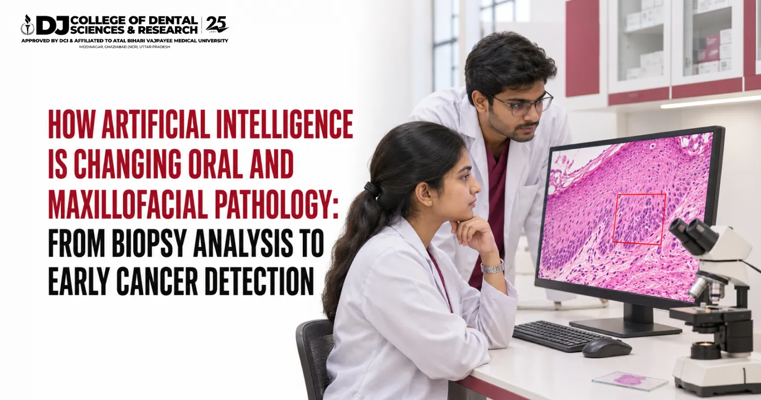
Artificial intelligence (AI) is changing oral maxillofacial pathology in three ways. It reads digital biopsy slides and marks suspicious cells. It grades pre-cancer changes with steadier results. It screens mouth photographs so that oral cancer is caught early. Pooled studies report about 90% sensitivity for AI in oral cancer diagnosis. The pathologist still gives the final report. India recorded 1,43,759 new oral cancer cases in 2022, so faster diagnosis matters here the most.
INTRODUCTION:
A small white patch inside the cheek looks harmless. Under a microscope, it can tell a very different story.
Reading that story is the daily work of oral maxillofacial pathology. For decades the tools were a glass slide, a microscope and a trained eye. The situation has changed considerably over the last few years. Software now scans the same slide and points to the cells that need a second look. This blog explains, in question-and-answer form, what AI does in the laboratory, how well it works and what it means for dental students.
What does an oral pathologist do?
• Oral maxillofacial pathology is the dental specialty that diagnoses diseases of the mouth, jaws and face.
• A biopsy is a small piece of tissue taken from a patch, ulcer or swelling.
• The pathologist stains it, studies it under a microscope and names the disease.
• The surgeon plans treatment only after this report arrives.
Why does India need faster oral cancer diagnosis?
India carries the heaviest oral cancer load in the world. Tobacco and areca nut account for about 70% to 80% of cases.
Table 1: Lip and oral cavity cancer in 2022
| Region | New cases | Deaths |
| World | 3,89,846 | 1,88,438 |
| India | 1,43,759 | 79,979 |
| China | 37,208 | 20,831 |
| United States | 27,750 | Not listed in top five |
| India’s share of the world total | About 37% | About 42% |
Source: GLOBOCAN 2022 figures published by the World Cancer Research Fund; shares calculated from the same figures.
The stage at diagnosis decides the outcome. Over half of Indian patients reach the hospital at stage III or IV.
Table 2: Five-year survival by stage at diagnosis
| Stage when cancer is found | Five-year survival |
| Early (stage I and II) | 70% to 90% |
| Advanced (stage III and IV) | 20% to 30% |
Source: International Journal of Community Medicine and Public Health; ecancermedicalscience GLOBOCAN 2022 analysis.
How is AI changing the pathology laboratory?
The first step is digital pathology. A scanner turns the glass slide into a whole-slide image, a very large digital picture of the tissue. AI then studies that picture.
Table 3: Traditional reporting and AI-assisted reporting
| Step | Traditional method | With AI support |
| Viewing the slide | Glass slide under a microscope | Digital slide on a screen |
| Finding abnormal areas | Pathologist scans every field by eye | Software highlights suspicious areas first |
| Grading pre-cancer | Depends on the reader; grades can differ | Same rule applied to every slide |
| Second opinion | Slide travels by post | Image shared online within minutes |
| Final report | Pathologist | Pathologist |
Source: Review of AI in digital pathology for oral epithelial dysplasia, Head and Neck Pathology (PMC11087425).
How does AI read a biopsy slide in oral maxillofacial pathology?
• The software cuts the whole-slide image into thousands of small tiles.
• A deep learning model checks each tile for odd cell shapes, crowded nuclei and broken tissue layers.
• It joins the tile results into a colour map and a risk score for the full slide.
• The pathologist reviews the map, agrees or disagrees, and writes the report.
This matters most for oral epithelial dysplasia, a pre-cancer change. Grading it is hard, and two experts can differ on the same slide. This is where things become interesting.
Table 4: What AI achieved on biopsy slides
| Study | Task | Result |
| ODYN pipeline (Communications Medicine, 2025) | Dysplasia or no dysplasia | F1-score 0.96 |
| ODYN pipeline (Communications Medicine, 2025) | Predicting which patch turns into cancer | AUROC 0.73, similar to pathologist grading |
| Deep learning model (npj Digital Medicine, 2025) | Predicting progression to cancer from 221 slides | 80% accuracy, AUROC 0.798 |
Source: Nature Portfolio journals, links given at the end. F1-score and AUROC are accuracy measures on a 0 to 1 scale.
Can AI detect oral cancer early?
Yes, as a screening aid. Two terms help here. Sensitivity is the share of diseased cases a test catches. Specificity is the share of healthy cases it clears correctly.
Table 5: Pooled accuracy of AI for oral cancer and pre-cancer
| Meta-analysis | Images studied | Sensitivity | Specificity |
| BMC Cancer, 2026 (all AI models) | Mixed | 90% | 89% |
| BMC Cancer, 2026 (image-based models) | Images | 94% | 93% |
| J Int Soc Prev Community Dent (35 studies, 2015 to 2024) | Mixed | 87% | 81% |
| Same study, clinical photographs only (21 studies) | Mouth photos | 86% | 78% |
Source: BMC Cancer 2026; Journal of International Society of Preventive and Community Dentistry.
What happened when AI was tested in Indian communities?
A field study in India screened 5,025 people with a phone-based device and collected 32,128 images.
Table 6: Who spotted suspicious mouth lesions best?
| Screener | Sensitivity | Specificity |
| Frontline health worker, by eye | 60% | 78% |
| AI model on the phone | 82% | 77% |
| AI model on a cloud server | 87% | Not stated here |
| Remote specialist viewing the images | 95% | 84% |
Source: Birur and others, Scientific Reports, 2022.
In practical terms, the phone model caught far more suspicious lesions than a health worker alone. The specialist still did best.
Will AI replace the oral pathologist?
No. Ask any experienced pathologist, and they will tell you that a slide is only one part of the diagnosis. AI in oral maxillofacial pathology has clear limits.
• Most studies use past records, not live patients.
• Results differ widely between studies, so one headline figure can mislead.
• In head-to-head tests, AI matched experts on sensitivity but tended to score lower on specificity.
• A model trained on one population may slip on another.
• Biopsy and microscopic study remain the gold standard for confirming cancer.
How big is the market for AI in pathology?
Table 7: Global AI in pathology market
| Year | Value in US dollars | Value in INR |
| 2025 | USD 168.3 million | ₹1,584 crore |
| 2026 (estimate) | USD 211.2 million | ₹1,988 crore |
| 2033 (forecast) | USD 1,151.6 million | ₹10,841 crore |
Source: Grand View Research; converted at the Reserve Bank of India rate of 6 November 2026 ($1 = ₹94.1402).
The forecast growth rate is 27.4% a year. Laboratories will need pathologists who can work with these tools.
Which oral maxillofacial course leads to this field?
Students often search for an oral maxillofacial course without knowing that two MDS branches carry the name. One diagnoses. The other operates.
Table 8: Two MDS branches at DJ College of Dental Sciences & Research
| Point | MDS Oral Pathology & Microbiology | MDS Oral & Maxillofacial Surgery |
| Main work | Diagnoses disease from biopsy tissue | Operates on the mouth, jaw and face |
| Duration | 3 years | 3 years |
| Entry | BDS, internship and NEET MDS | BDS, internship and NEET MDS |
| MDS seats | 6 | 3 |
| Tuition per year (2026) | ₹2,00,000 | ₹9,25,000 |
| Link with AI | Reads AI-marked slides and signs the report | Uses the report to plan surgery |
Source: DJ College website; fee details filed with DGME UP for 2026, as reported by Medical Dialogues.
Oral maxillofacial pathology at DJ College of Dental Sciences & Research, Modinagar
• The college stands in Modinagar, Ghaziabad district, within Delhi NCR, and has taught dentistry since 1999.
• The Department of Oral Pathology & Microbiology works beside eight other departments, so biopsy cases come from every clinic.
• 100+ patients visit each department every day on a 75-acre campus.
• The Dental Council of India recognises its BDS and MDS courses, and students come from 17+ countries.
• Research and publication form part of MDS training, which suits a field moving towards digital slides.
What does an oral maxillofacial career in 2027 look like?
An oral maxillofacial career 2027 onwards will mix microscope skill with screen skill.
• Diagnostic pathologist in hospitals and private laboratories.
• Teacher in dental colleges, where every BDS student studies oral pathology.
• Screening specialist in oral cancer programmes that use phone-based tools.
• Researcher who trains and tests AI models on Indian slides.
• Consultant to digital pathology companies.
Conclusion
AI has given oral maxillofacial pathology a second pair of eyes. It scans slides quickly, grades pre-cancer with steadier rules and helps health workers catch suspicious patches early. It does not sign the report. The pathologist does.
For a country with 1,43,759 new oral cancer cases a year, that partnership can save lives. For a BDS graduate, it opens a specialty where laboratory skill and computer skill meet. DJ College of Dental Sciences & Research, Modinagar, trains MDS students in both Oral Pathology & Microbiology and Oral & Maxillofacial Surgery on one campus.
This article is for education only. It is not medical advice. Anyone with a mouth patch or ulcer that lasts over two weeks should see a dentist.
Frequently Asked Questions (FAQs)
1. What is oral and maxillofacial pathology?
It is the dental specialty that finds the cause of diseases of the mouth, jaws and face. The oral pathologist studies biopsy tissue under a microscope and gives the final diagnosis.
2. How is artificial intelligence used in oral pathology?
AI software reads digital images of biopsy slides and mouth photographs. It marks suspicious areas, helps grade pre-cancer changes and sorts urgent cases for the pathologist.
3. Can AI detect oral cancer early?
Studies say it can help. One 2026 meta-analysis found pooled sensitivity of 90% and specificity of 89% for AI models in oral cancer diagnosis. A biopsy is still needed to confirm.
4. Will AI replace oral pathologists?
No. AI works as a second reader. The pathologist checks the result, weighs the patient history and signs the report.
5. How many oral cancer cases does India report?
GLOBOCAN 2022 recorded 1,43,759 new lip and oral cavity cancer cases and 79,979 deaths in India, the highest of any country.
6. Why does early detection of oral cancer matter?
Five-year survival is about 70% to 90% when oral cancer is found in stage I or II. It drops to about 20% to 30% in advanced stages.
7. What is a whole-slide image?
It is a high-resolution digital scan of a full glass biopsy slide. AI needs this scan to study the tissue.
8. Which course leads to a career in oral pathology?
MDS in Oral Pathology & Microbiology, a three-year course after BDS. Admission is through NEET MDS and counselling.
9. Does DJ College of Dental Sciences & Research offer MDS in Oral Pathology?
Yes. DJ College, Modinagar, has 6 MDS seats in Oral Pathology & Microbiology and 3 in Oral & Maxillofacial Surgery, recognised by the Dental Council of India.
10. What jobs can an oral pathologist take up in 2027?
Common roles are diagnostic laboratory pathologist, dental college teacher, cancer screening programme specialist, researcher and consultant to digital pathology firms.
Related Posts