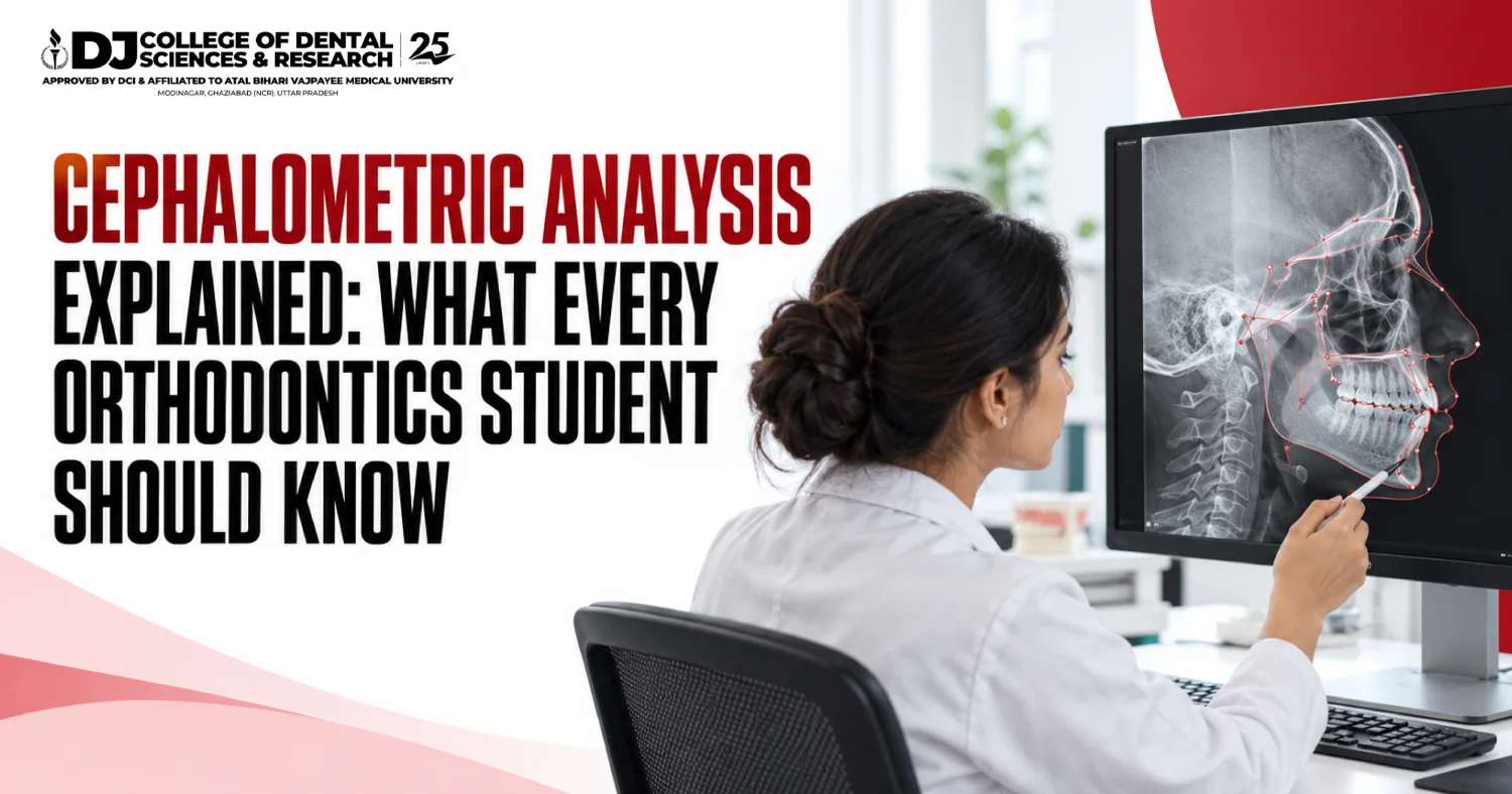
Cephalometric analysis is the study of a side-view X-ray of the head. It measures how the jaws, teeth and face relate to each other. Students mark fixed points, join them into lines and read the angles. Three angles do most of the early work: SNA (82 degrees), SNB (80 degrees) and ANB (2 degrees). The best dental colleges in India teach it by hand first and on software later.
A third-year BDS student sits in front of a light box. On it lies a grey side-view X-ray of a stranger’s skull, a sheet of tracing paper and a sharp pencil. The teacher says, “Mark Sella.” The student stares. Nothing on the film looks like a saddle.
For many students, this is the first real meeting with orthodontics, and it is rarely love at first sight. Yet this one film quietly decides big things. It tells whether a child needs braces or a growth appliance. It tells whether an adult needs jaw surgery.
That is why the best dental colleges in India usually ask students to trace by hand before they touch any software. The skill looks dull. It turns out to be the backbone of diagnosis.
This blog from DJ College of Dental Sciences & Research, Modinagar, explains the subject in plain words: the points, the lines, the famous angles, the safety numbers and the courses that use it.
What Is Cephalometric Analysis in Simple Words?
• A cephalogram is a side-view X-ray of the skull, taken in a head holder called a cephalostat.
• The X-ray source sits about five feet from the middle of the head, and the film about 15 cm away. This fixed set-up makes every film comparable.
• Cephalometric analysis means measuring that film to judge the jaws, teeth and facial profile.
Who Started It, and Why Does It Still Matter?
Holly Broadbent Sr. introduced the cephalostat in the USA in 1931. Cecil Steiner followed with his famous analysis in 1953. Nearly a century on, orthodontists still use it to:
• find out whether the fault lies in the jaw or in the teeth
• judge the direction of a child’s growth
• plan braces, tooth removal or jaw surgery
• compare the face before and after treatment
Which Landmarks Should Every Orthodontics Student Know First?
Landmarks are fixed points on the skull. Everything else hangs on them.
| Landmark | Short form | Where it sits |
| Sella | S | Centre of the bony saddle at the base of the skull |
| Nasion | N | Front point where the forehead meets the nose bones |
| A point | A | Deepest point on the front of the upper jaw |
| B point | B | Deepest point on the front of the lower jaw |
| Pogonion | Pg | Most forward point of the chin |
| Menton | Me | Lowest point of the chin |
| Gonion | Go | Back lower corner of the lower jaw |
| Orbitale | Or | Lowest point on the rim of the eye socket |
| Porion | Po | Top of the ear canal opening |
Which Lines Join These Points?
• SN plane: Sella to Nasion. It stands for the front of the skull base.
• Frankfort horizontal: Porion to Orbitale.
• Mandibular plane: along the lower border of the lower jaw.
What Do SNA, SNB and ANB Mean?
This is where things become interesting. A few angles turn a grey film into a diagnosis.
| Measurement | Normal value | What it shows |
| SNA | 82 degrees (plus or minus 2) | Position of the upper jaw |
| SNB | 80 degrees (plus or minus 2) | Position of the lower jaw |
| ANB | 2 degrees (plus or minus 2) | Gap between the two jaws |
| FMA (Tweed) | 25 degrees | Steepness of the lower jaw |
| IMPA (Tweed) | 90 degrees (plus or minus 5) | Tilt of the lower front teeth |
| Wits | -1 mm men, 0 mm women | Jaw relation along the bite plane |
• ANB near 2 degrees suggests a Class I (normal) jaw pattern.
• ANB of 4 degrees or above suggests Class II, where the lower jaw sits behind.
• ANB of zero or below suggests Class III, where the lower jaw sits ahead.
Which Analyses Do the Best Dental Colleges in India Teach First?
• Steiner (1953) and Downs, the two most popular methods.
• Tweed (1966), built around the lower front teeth.
• Wits (1975), a quick check on the jaw relation.
• Jarabak (1972) and Ricketts, for growth pattern and facial form.
No single analysis tells the whole story. Teachers at the top dental colleges in India ask students to read two or three together.
How Do the Best Dental Colleges in India Move Students From Pencil to Software?
• Hand tracing first. It trains the eye to find each point.
• Digital tracing next. Software measures the angles in seconds.
• AI tools last. A 2021 review of 19 studies found that deep learning placed 79.9% of points within 2 mm. The authors also warned of a high risk of bias, so a trained person must still check every point.
Ask any experienced professional in the field, and the reply stays the same: the sums are easy, finding the points is hard.
Is a Cephalogram Safe, and What Do the Numbers Say?
| Fact | Figure | Source |
| Radiation from one lateral cephalogram | 5.03 microsieverts | J Orofac Orthop, 2016 |
| Radiation from one panoramic X-ray | 21.87 microsieverts | J Orofac Orthop, 2016 |
| Radiation from one CBCT scan (portrait mode) | 131.7 microsieverts | J Orofac Orthop, 2016 |
| BDS seats in India, 2026 | About 28,000 in 329 colleges | Vedantu, citing DCI and NDC data |
| MDS seats in India, 2025 | About 6,228 in 259 colleges | Amar Ujala |
A cephalogram carries a small dose, far below a 3D scan. The imaging business behind it keeps growing too.
| Global dental imaging market | Figure in US dollars | In Indian rupees (INR) |
| Size in 2025 | USD 3.8 billion | About Rs 36,784 crore |
| Forecast for 2034 | USD 6.9 billion | About Rs 66,792 crore |
Converted at Rs 96.8 to one US dollar, the rate on 8 October 2026.
Which Dental Courses and Careers Use Cephalometric Analysis?
Not everyone is aware that this skill travels across several dental courses and dental specialisations.
| Course | Duration | How it uses cephalometrics |
| BDS | 4 years + 1 year internship | Basics of tracing in orthodontics |
| MDS Orthodontics & Dentofacial Orthopedics | 3 years | Daily diagnosis and treatment planning |
| MDS Oral & Maxillofacial Surgery | 3 years | Planning jaw operations |
| MDS Oral Medicine & Radiology | 3 years | Taking and reading skull X-rays |
A dental career built on it can lead to work as an orthodontist, a jaw surgeon, an oral radiologist, a teacher or a researcher.
How Does DJ College of Dental Sciences & Research Teach It?
Many parents and students still believe that the best dental colleges in India are only the names at the top of a ranking list. The reality on the ground is somewhat different. What a student handles in the clinic counts just as much. The Department of Orthodontics & Dentofacial Orthopedics at DJ College offers:
• Dolphin software and CBCT for diagnosis.
• A postgraduate clinic with 22 dental chairs.
• 1,000+ orthodontic patients a month, so students see many kinds of cases.
• Orthodontic training from the third year of BDS, under close supervision.
• A three-year MDS in Orthodontics with six seats.
• A 75-acre campus in Modinagar, Ghaziabad, serving Delhi NCR since 1999, with students from 17+ countries.
• Recognition by the Dental Council of India (DCI), now succeeded by the National Dental Commission (NDC).
Readers comparing the top dental colleges in India can run the same checks on any campus.
Conclusion: Why Does One Side-View X-Ray Matter So Much?
Cephalometric analysis turns a face into numbers that a dentist can trust. A student who can find Sella, draw the SN plane and read ANB already thinks like an orthodontist.
Software and AI will keep getting faster, but they still need a trained eye behind them. That eye grows in clinics with real patients and patient teachers, which is what the best dental colleges in India have in common. Students who want to train this way can contact the admissions team at DJ College of Dental Sciences & Research, Modinagar.
Frequently Asked Questions
1. What is cephalometric analysis?
Cephalometric analysis is the study of a side-view X-ray of the head. It measures how the jaws, teeth and face relate, so that orthodontists can plan treatment.
2. What is a cephalogram?
A cephalogram is a standard side-view skull X-ray taken in a head holder called a cephalostat. The fixed set-up makes films comparable over time.
3. What are SNA, SNB and ANB?
They are three angles from the Steiner analysis. SNA shows the position of the upper jaw, SNB the lower jaw, and ANB the gap between the two.
4. What is a normal ANB angle?
A normal ANB angle is about 2 degrees. A value of 4 degrees or above points to a Class II pattern, and zero or below points to Class III.
5. Who introduced cephalometrics?
Holly Broadbent Sr. introduced the cephalostat in the USA in 1931. Cecil Steiner published his well-known analysis in 1953.
6. Is a cephalometric X-ray safe?
The dose is small. One study measured 5.03 microsieverts for a lateral cephalogram, against 21.87 for a panoramic X-ray.
7. Can AI do cephalometric tracing?
AI tools can place the points quickly. A 2021 review of 19 studies found 79.9% of points within 2 mm, so a trained person must still check them.
8. In which year do BDS students learn cephalometric analysis?
Orthodontics is taught in the later years of BDS. At DJ College of Dental Sciences & Research, orthodontic training starts in the third year.
9. Which dental specialisations use cephalometric analysis?
Orthodontics uses it daily. Oral and maxillofacial surgery uses it to plan jaw operations, and oral radiology and children’s dentistry use it too.
10. Which are the best dental colleges in India for orthodontics?
No single list settles it. Students should look for heavy patient flow, digital tracing software, CBCT and teachers who check every tracing.
Related Posts